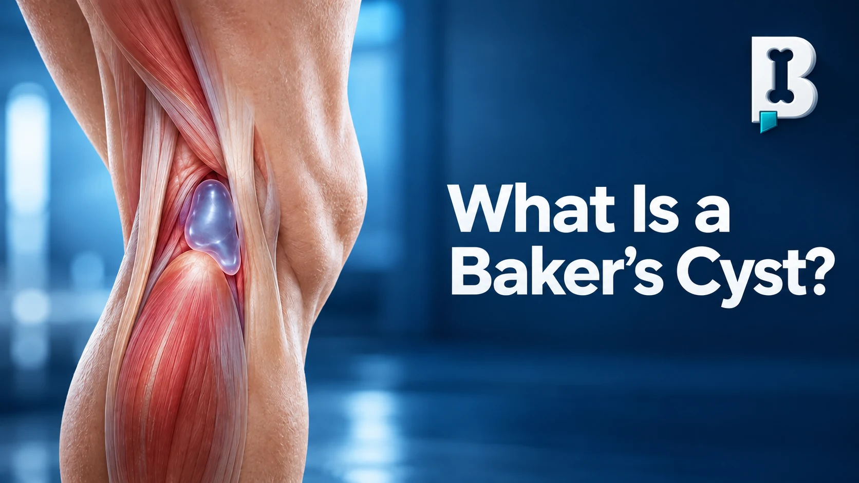
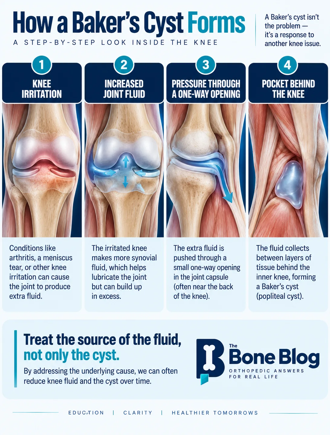
Think of squeezing a water balloon between your hands. Pressure makes a small pouch bulge through the opening between your fingers. In the knee, fluid can move through a communicating area in the posterior capsule and collect between the medial head of the gastrocnemius and the semimembranosus tendon.
Why Does It Form?
The knee normally contains synovial fluid. Irritation inside the joint increases fluid production and pressure. The posterior communication can function like a one-way valve, allowing fluid into the gastrocnemius-semimembranosus bursa but limiting its return. The fluid may become thick or jelly-like as the pocket enlarges.
This is why I tell patients that a Baker’s cyst is often a symptom of the knee’s underlying problem rather than a separate disease that needs to be chased.
What Does It Feel Like?
Many Baker’s cysts cause no symptoms and are found incidentally on imaging. When symptomatic, they may cause fullness or tightness behind the inner side of the knee, sometimes with limited bending. Pain often comes from arthritis or a meniscus tear that produced the fluid.
How Is It Diagnosed?
History and examination come first. As one of my residency attendings said, “When all else fails, examine the patient.” A lump behind the knee should not automatically be labeled a Baker’s cyst.
Other possibilities include a lipoma, another fluid collection, tendon injury, a bony prominence, a blood-vessel problem, or, less commonly, a solid mass. Tenderness, fixation, skin changes, rapid growth, or an unusual location make further evaluation more important.
X-rays help evaluate arthritis, alignment, and bone. Ultrasound can confirm that a mass is fluid-filled and evaluate the veins when a blood clot is possible. MRI is most useful when assessing the cyst along with a meniscus tear, cartilage damage, or other intra-articular pathology.
What If It Ruptures?
A cyst can leak and let fluid track down the calf. Patients sometimes describe water running down the back of the leg. The fluid is usually reabsorbed, but sudden calf swelling or pain can resemble deep vein thrombosis. New calf swelling, warmth, redness, substantial tenderness, chest pain, or shortness of breath requires prompt evaluation.
How Is It Treated?
A painless incidental cyst usually needs education and observation, not a procedure. Compression with an elastic wrap or sleeve may reduce mild fullness.
Treatment should focus on why the knee is producing excess fluid. Depending on the diagnosis, options may include rehabilitation, medication, aspiration of a large knee effusion, or an intra-articular injection. Corticosteroid, viscosupplementation, and orthobiologic injections are not interchangeable, and the choice depends on the underlying condition.
I do not routinely aspirate the cyst itself. Ultrasound-guided aspiration and corticosteroid injection may help selected symptomatic cysts, but recurrence is possible if the knee keeps producing fluid or the valve remains.
When Is Surgery Considered?
Surgery is uncommon. It may be considered when a clearly symptomatic cyst persists despite nonsurgical care and treatment of the underlying knee problem. Large or complicated cysts causing nerve or blood-vessel compression need more urgent attention.
Multiloculated cysts contain internal partitions and can be difficult to drain. Persistent arthritis, capsular weakness, or the valve mechanism can allow a cyst to recur even after aspiration or removal.
A Patient Example
One patient came to me because they had been told they had a Baker’s cyst. There was fullness behind the knee, but the X-rays and history pointed much more strongly to symptomatic arthritis. We treated the arthritis. At follow-up, when I asked about the cyst, the patient replied, “What Baker’s cyst?” The named finding had dominated the initial concern, but it was a sign of the problem rather than the problem itself.
When Should You Be Evaluated?
A stable, painless cyst found incidentally is rarely urgent. Arrange evaluation if fullness is painful, persistent, enlarging, limiting motion, or accompanied by mechanical knee symptoms. Seek faster assessment for rapid enlargement, significant pain, warmth, redness, skin changes, calf swelling, new instability, major motion loss, numbness, or weakness. Chest pain or shortness of breath is an emergency.
Bottom Line
Most Baker’s cysts are not dangerous and do not need removal. The better question is not only how to get rid of the cyst, but why the knee is producing extra fluid. Treating the arthritis, meniscus problem, or inflammation usually matters more than treating the lump.
References
- Frush TJ, Noyes FR. Baker’s Cyst: Diagnostic and Surgical Considerations. Sports Health. 2015.
- Van Nest DS, et al. Popliteal Cysts: A Systematic Review of Nonoperative and Operative Treatment. JBJS Reviews. 2020.
- Smith MK, et al. Treatment of Popliteal (Baker) Cysts With Ultrasound-Guided Aspiration, Fenestration, and Injection: Long-term Follow-up. Sports Health. 2015.
Visual summary
Frequently asked questions
Is a Baker’s cyst dangerous?
Most Baker’s cysts are not dangerous. They are usually a sign that the knee is producing extra fluid because of arthritis, a meniscus problem, cartilage damage, or inflammation.
Can a Baker’s cyst rupture?
Yes. Fluid can leak down the calf and cause sudden pain or swelling. Because this can resemble a blood clot, new calf swelling, warmth, redness, or significant tenderness should be evaluated promptly.
Does a Baker’s cyst need to be drained?
Not usually. Aspiration can help selected symptomatic cysts, but recurrence is common when the knee continues to make excess fluid. Treatment usually focuses on the underlying knee problem.
Can arthritis cause a Baker’s cyst?
Yes. Arthritis is a common reason the knee produces extra synovial fluid, which can then collect behind the knee as a Baker’s cyst.
Can a meniscus tear cause a Baker’s cyst?
Yes. Meniscus irritation or tearing can contribute to inflammation and excess joint fluid, which may promote formation of a Baker’s cyst.
When should a Baker’s cyst be evaluated?
Arrange evaluation if the fullness is painful, persistent, enlarging, limiting motion, or accompanied by mechanical symptoms. Seek faster assessment for rapid growth, warmth, redness, major calf swelling, numbness, weakness, or significant loss of motion.
Need a joint checked?
If pain, instability, an injury, or mechanical symptoms are limiting your activity, an orthopedic evaluation can help clarify the cause.
Schedule an Appointment