
An X-ray is often a reasonable first step when you have had a meaningful injury, cannot comfortably bear weight, notice a deformity, or have pain that is not improving with rest and activity modification. It is an excellent screening tool, but it is only one part of the evaluation.
Your symptoms, the mechanism of injury, your medical history, and a physical examination all help determine whether an X-ray is needed and what imaging—if any—should come next.
What can an X-ray show?
X-rays are one of the standard first-line tests used in orthopedics because they provide a quick way to evaluate bones and joints. They can help identify fractures, dislocations or partial dislocations, arthritis and loss of joint space, changes in bone alignment, old healed injuries, certain abnormalities in bone quality, and changes in the position of a known fracture over time.
I have reviewed well over a million X-rays during my career, and they often tell a story. An image may show the immediate injury, but it may also reveal an old healed fracture, longstanding arthritis, altered alignment, or clues about the quality of the bone.
Before becoming a doctor, I worked as a bank teller. We were taught to recognize counterfeit currency not by memorizing every possible counterfeit, but by learning exactly how authentic currency looked and felt. X-rays are similar. When you know normal anatomy extremely well, abnormalities often stand out.
When is an X-ray especially reasonable?
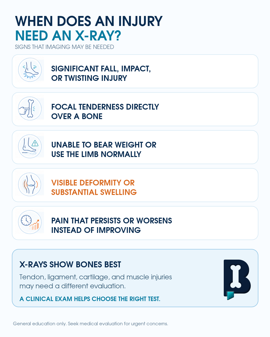
An X-ray may be appropriate after a fall, collision, twisting injury, or direct blow; when standing or bearing weight is difficult; when pain is focused directly over a bone; when there is significant swelling or bruising after trauma; when there is visible deformity or loss of normal motion; when pain is persistent rather than improving; when arthritis, instability, or abnormal alignment is suspected; or when a known fracture needs follow-up.
Why are several X-ray views needed?
In orthopedics, we often say, “One view is no view.” An X-ray turns a three-dimensional body part into a two-dimensional image. A fracture or alignment problem that is difficult to see from one direction may become obvious from another.
For that reason, we commonly obtain at least two views, such as a front view and a side view. Depending on the joint or suspected injury, additional angled, standing, weight-bearing, or stress views may be needed.
For example, non-weight-bearing hip or knee X-rays may underestimate arthritis because the joint is not being loaded normally. Standing views may reveal joint-space narrowing that was not apparent on the original images.
Can an X-ray be normal even when something is wrong?
Yes. A normal X-ray does not necessarily mean that nothing is injured. Standard X-rays do not clearly show many ligaments, tendons, menisci, cartilage injuries, muscle injuries, or some subtle and nondisplaced fractures.
Certain fractures may also be difficult to see initially because of their position, the angle of the images, or overlapping bones. When symptoms and examination findings remain concerning, repeat or specialized X-ray views, CT, or MRI may be appropriate.
A CT scan is often useful when more detail is needed about cortical bone, subtle fracture lines, displacement, or a complex fracture pattern. MRI is generally more helpful for ligaments, tendons, cartilage, bone marrow injuries, stress injuries, and other soft-tissue conditions.
Do soft-tissue injuries need repeat X-rays?
Usually not. Once adequate X-rays show no bony injury and the history and examination point toward a soft-tissue condition, repeating the same X-rays often provides little additional information. Repeat radiographs may become reasonable if there is a new injury, a significant change in symptoms, or concern that an initially subtle fracture has become more visible.
Follow-up X-rays are also commonly used to determine whether a known fracture has shifted, displaced, or changed alignment during healing. Those changes may affect the recommended treatment.
How long should I wait before getting an X-ray?
There is no single timeline that applies to every patient. After a substantial injury, immediate imaging may be appropriate. In other situations, a patient may initially try rest, ice, compression, elevation, and activity modification.
Pain tolerance also varies. A fracture that causes severe pain in one person may cause more manageable symptoms in another. The fact that someone can tolerate the pain does not necessarily rule out an injury.
Persistent pain that is not improving—particularly after a reasonable injury mechanism—deserves an evaluation.
Are X-rays safe?
Modern plain X-rays generally use relatively low radiation doses. When an X-ray is ordered to answer a meaningful clinical question, the expected benefit usually outweighs the small radiation exposure.
CT scans use more radiation than standard radiographs, which is one reason they are selected more carefully. However, CT can still be the correct test when detailed three-dimensional information about a fracture is needed.
The goal is not to avoid all imaging. It is to select the study that is most likely to answer the clinical question without ordering unnecessary tests.
How I evaluate this in clinic
I begin by asking how the pain started, where it is located, what makes it worse, and whether the patient can use or bear weight on the injured area. I then examine the joint or extremity for focal tenderness, swelling and bruising, alignment, range of motion, instability, strength, and neurovascular function.
I interpret the X-rays alongside that history and examination. Each piece is part of the puzzle. From there, I decide whether the patient needs conservative treatment, additional X-ray views, CT, MRI, immobilization, follow-up imaging, or another form of care.
When should you seek an evaluation?
Consider an in-person evaluation when pain follows a significant injury, you cannot use the limb normally, weight-bearing is difficult, there is visible deformity, or symptoms are persistent rather than steadily improving.
An X-ray may be an excellent place to start, but the image should not be interpreted in isolation. The best decisions come from putting the X-ray together with the patient’s story and a careful physical examination.
This article provides general orthopedic education and is not a diagnosis or individualized medical recommendation.
References
- Taljanovic MS, et al. ACR Appropriateness Criteria Acute Trauma to the Knee. Journal of the American College of Radiology. 2020.
- Stiell IG, et al. Prospective Validation of a Decision Rule for the Use of Radiography in Acute Knee Injuries. JAMA. 1996.
- Bachmann LM, et al. Accuracy of Ottawa Ankle Rules to Exclude Fractures of the Ankle and Mid-foot: Systematic Review. BMJ. 2003.
Visual summary
Frequently asked questions
Does every injury need an X-ray?
No. Minor injuries that are steadily improving may not require imaging. The mechanism of injury, location of tenderness, ability to use the extremity, and examination findings all help determine whether an X-ray is appropriate.
Can an X-ray show a ligament or tendon tear?
Usually not directly. X-rays primarily show bones, joint spacing, and alignment. MRI or ultrasound may be more useful for suspected soft-tissue injuries.
Can I still have a fracture if the first X-ray is normal?
Yes. Some subtle, nondisplaced, stress, or anatomically obscured fractures may not be visible initially. Repeat X-rays, CT, or MRI may be needed when symptoms and examination remain concerning.
Why does my doctor order several X-ray views?
Each view shows the anatomy from a different direction. A fracture, dislocation, or alignment problem may be hidden on one image and clearer on another.
Why might I need standing or weight-bearing X-rays?
They show how a joint behaves under normal load and can reveal joint-space narrowing, arthritis, instability, or alignment abnormalities that may not be apparent on a non-weight-bearing image.
Are repeated X-rays dangerous?
The radiation dose from standard plain radiographs is generally low. Imaging should still be ordered thoughtfully, but repeated X-rays may be appropriate to diagnose an injury or monitor fracture alignment and healing.
Need a joint checked?
If pain, instability, an injury, or mechanical symptoms are limiting your activity, an orthopedic evaluation can help clarify the cause.
Schedule an Appointment