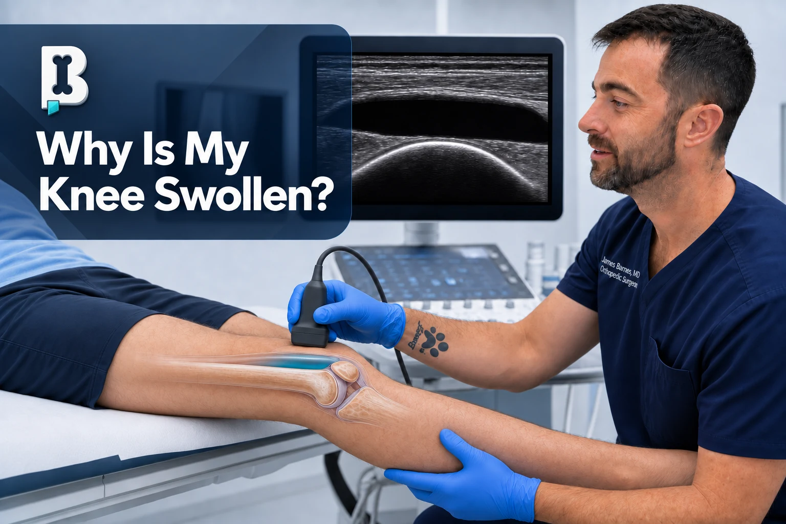
A swollen knee is a symptom, not a diagnosis. The swelling may represent extra lubricating fluid inside the joint, blood from an acute injury, inflammation around the knee, crystal irritation such as gout, or—less commonly but more urgently—an infection.
The timing, location, associated symptoms, injury history, examination, imaging, and sometimes a sample of the fluid help determine what is causing it.
The first questions matter
When I evaluate a swollen knee, I start by building the clinical picture:
- How long has the swelling been present?
- Has it happened before?
- Was there an injury?
- Did it appear immediately, later that day, or gradually over weeks?
- Is the swelling diffuse or concentrated in one spot?
- Does it seem to be inside the joint or outside it?
- Is the knee red, hot, or severely painful?
- Can the patient bear weight?
- Is it a native knee or a knee replacement?
- Has the knee had prior surgery, a recent injection, or another procedure?
- What makes the swelling better or worse?
Those details often narrow the possibilities before advanced imaging is ever considered.
Is the fluid inside or outside the knee joint?
Not every swollen-looking knee contains a true joint effusion.
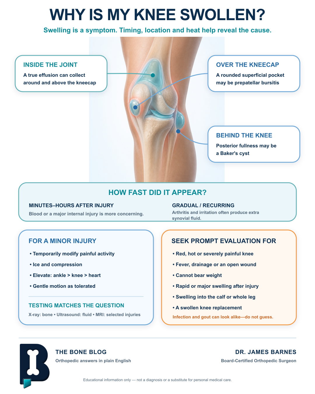
A prepatellar bursitis often looks like an egg sitting directly over the kneecap. Fullness behind the knee may suggest a Baker’s cyst. Swelling may also be located in the soft tissues outside the joint capsule or may extend from the calf or lower leg.
A true effusion is fluid inside the knee joint. With the knee straight, an examiner may be able to gather the fluid beneath the kneecap and make the patella feel as though it is floating. This is sometimes called a ballotable patella. Ultrasound can also confirm whether fluid is present and help identify where it is located.
The amount, shape, location, tenderness, redness, and warmth of the swelling all provide useful clues.
What kind of fluid can collect in a knee?
Several types of fluid can be found inside a swollen knee.
Synovial fluid. Synovial fluid normally lubricates the joint and helps the cartilage surfaces glide. When an arthritic knee becomes irritated, the synovial lining may produce more fluid. That increased production can create the recurring swelling patients often describe as “fluid on the knee.”
Blood. Rapid swelling within minutes to hours after an injury may represent bleeding into the joint. This can occur when blood vessels or internal structures are disrupted. A classic example is a noncontact pivoting injury with a pop and immediate swelling, which raises concern for an ACL injury or another significant internal derangement.
Inflammatory or crystal-containing fluid. Gout and pseudogout can cause a knee to become suddenly red, hot, swollen, and very painful. These conditions can resemble infection. Examining the joint fluid under a microscope may reveal crystals and help establish the diagnosis.
Purulent fluid. An infected joint may contain cloudy, yellow-white, or pus-like fluid. Appearance alone is not enough to make or exclude the diagnosis, so the sample generally needs laboratory analysis.
Why does arthritis make a knee swell?
Arthritic swelling often reflects a balance between activity and inflammation. When the joint is irritated, the synovial lining may produce more fluid in an attempt to improve lubrication and cushioning.
That does not mean the answer is complete inactivity. Motion helps preserve mobility, strength, and function—what many clinicians mean when they say, “motion is lotion.” The goal is to remain active at a level the knee can tolerate rather than repeatedly pushing it into a flare.
Anti-inflammatory treatments may reduce the inflammatory response and, in turn, reduce excess synovial-fluid production. Depending on the patient and the cause, this may include an oral nonsteroidal anti-inflammatory drug or a corticosteroid injection. These treatments are not appropriate for everyone and should be considered in the context of the patient’s medical history.
How do acute injuries cause swelling?
Most people remember a meaningful acute knee injury. The mechanism may be a pivot, twist, direct impact, fall, or sudden change of direction. The faster and more dramatically the knee swells, the more concerned I become about a significant intra-articular injury.
The history may be more informative than the examination at first. A painful, guarded knee may not tolerate every provocative maneuver. In that setting, I do as much of the examination as is reasonable, consider the mechanism, obtain appropriate imaging, and reassess as pain and swelling permit.
Mechanical symptoms such as instability, true locking, catching, or joint-line pain may add useful information, but they must be interpreted together with the entire clinical picture.
When could a swollen knee be infected?
A knee infection requires prompt evaluation. Features that raise concern include:
- Increasing redness, warmth, swelling, or severe pain
- Significant pain with motion or weight bearing
- Fever or recent systemic illness
- Drainage or an open wound
- Recent surgery, injection, or other procedure
- A knee replacement or other implanted hardware
- Diabetes, immune suppression, or a weakened immune response
- Skin breakdown near the knee
The skin is the body’s best natural barrier. If that barrier is compromised, bacteria may have a route into deeper tissues. Bacteria can also occasionally reach the joint through the bloodstream after another illness.
Infection and crystal arthritis can look very similar. A red, hot, swollen knee is therefore not something to diagnose by appearance alone.
When is knee aspiration helpful?
Aspiration, also called arthrocentesis, means placing a needle into the joint and removing fluid. It may be diagnostic, therapeutic, or both.
The fluid’s appearance can provide immediate clues. Bright red blood may follow an injury. Amber-colored fluid is commonly seen with osteoarthritis. Cloudy or pus-like fluid raises concern for infection. However, visual appearance is not definitive.
Laboratory analysis may include a cell count, crystal analysis, Gram stain, and culture. These tests help identify inflammation, gout or pseudogout, and possible infection.
Ultrasound can confirm that fluid is actually present and help guide the needle into the joint. When little or no fluid is visible, an attempted aspiration may result in a “dry tap.”
Which imaging test is best?
The right imaging test depends on the question being asked.
X-rays are often the first test after an injury. They are useful for identifying fractures, alignment problems, arthritis, and other bony changes.
Ultrasound can localize fluid, distinguish some superficial fluid collections from a true joint effusion, evaluate selected structures dynamically, and guide aspiration or injection. It cannot see through bone, so it has limitations for deeper structures inside the knee.
MRI provides detailed views of structures that are difficult to assess with ultrasound, including the menisci, ACL, PCL, cartilage, bone marrow, and other internal tissues. It is most useful when the examination and initial imaging do not adequately explain the swelling or when a specific internal injury is suspected.
MRI is not automatically the first or best test for every swollen knee. The imaging should answer a specific clinical question.
What can I do at home?
For swelling after a relatively minor injury, let pain be your guide. One practical rule is that if pain remains increased for more than about an hour after stopping an activity, you probably exceeded what the knee currently tolerates and should back off.
Early home care may include:
- Rest or temporary activity modification
- Ice
- Compression
- Elevation
- Gentle motion as tolerated
For meaningful elevation, place the ankle above the knee and the knee above the heart. Fluid follows gravity, so simply resting the foot on a low stool may not substantially reduce swelling.
Over-the-counter anti-inflammatory medications such as ibuprofen or naproxen may help some patients. They are not safe for everyone, including certain people with kidney disease, medication interactions, bleeding risk, or other medical conditions. Follow the product instructions and ask a clinician or pharmacist when uncertain.
Trying to push through an acutely swollen, painful knee can increase irritation and produce even more swelling.
When should I get the knee evaluated?
Seek prompt or urgent evaluation when the knee is becoming worse rather than better, especially with:
- A red, hot, severely painful knee
- Fever or drainage
- An open wound or compromised skin
- Obvious deformity
- Inability to bear weight
- Rapid or major swelling after an injury
- Swelling extending into the calf or throughout the leg
- Significant pain with even gentle motion
- A swollen knee replacement
- Symptoms that are not progressively improving with reasonable home care
Calf or whole-leg swelling may come from something other than the knee itself and can require a different evaluation.
It is also appropriate to be examined simply because you are unsure. Identifying the exact cause of swelling is not the patient’s job. Your job is to listen to your body, notice whether the condition is improving or worsening, and seek help when something does not seem right. The physician’s job is to assess, diagnose, and treat the underlying cause.
The bottom line
Knee swelling may come from arthritis, an acute injury, bursitis, a Baker’s cyst, crystal arthritis, infection, or another source. The most useful clues are how quickly it started, whether an injury occurred, where the swelling is located, whether the knee is hot or red, and whether you can comfortably move it and bear weight.
A swollen knee that is improving may respond to temporary activity modification, ice, compression, elevation, and gentle motion. A knee that is increasingly red, hot, painful, difficult to use, draining, or associated with fever deserves prompt medical attention—especially when a knee replacement is present.
This article provides general medical education and does not diagnose an individual condition or replace an in-person examination. Seek urgent care for severe or rapidly worsening symptoms, fever, drainage, inability to bear weight, deformity, or concern for infection.
References
- Duong V, et al. Evaluation and Treatment of Knee Pain: A Review. JAMA. 2023.
- Margaretten ME, et al. Does This Adult Patient Have Septic Arthritis? JAMA. 2007.
- Dalbeth N, et al. Gout. The Lancet. 2021.
- Taljanovic MS, et al. ACR Appropriateness Criteria Acute Trauma to the Knee. Journal of the American College of Radiology. 2020.
Visual summary
Frequently asked questions
Is a swollen knee always something to worry about?
Not always, but it is worth paying attention to. A gradual, recurrent effusion often comes from arthritis, while a knee that is becoming worse rather than better — especially if it is red, hot, severely painful, draining, or associated with fever — needs prompt evaluation.
What is the difference between fluid inside the knee and swelling around it?
A true effusion is fluid inside the joint itself and can make the kneecap feel like it is floating. Swelling outside the joint, such as a prepatellar bursitis, often looks like a distinct egg-shaped lump directly over the kneecap rather than a diffuse fullness.
Can arthritis cause a knee to swell?
Yes. When an arthritic joint is irritated, the synovial lining may produce more lubricating fluid, which can create recurring swelling. Staying appropriately active while avoiding repeated flares, along with anti-inflammatory treatment when appropriate, can help.
When does a swollen knee need urgent evaluation?
Seek prompt care for a red, hot, severely painful knee, fever or drainage, an open wound, obvious deformity, inability to bear weight, rapid swelling after an injury, swelling spreading into the calf or leg, or a swollen knee replacement.
What can I do at home for minor knee swelling?
For swelling after a relatively minor injury, rest or activity modification, ice, compression, and elevation with the ankle above the knee and the knee above the heart may help. If the knee is not progressively improving, it should be evaluated.
Need a joint checked?
If pain, instability, an injury, or mechanical symptoms are limiting your activity, an orthopedic evaluation can help clarify the cause.
Schedule an Appointment