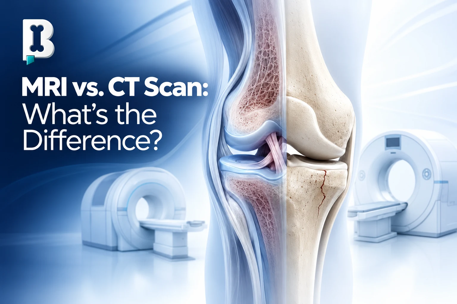
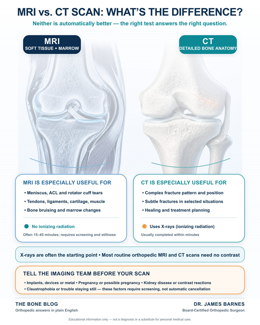
An MRI and a CT scan are not the same test, and neither one is automatically better. In orthopedics, CT is especially useful for detailed views of bone, while MRI is especially useful for soft tissues such as tendons, ligaments, cartilage, and muscles. The right study depends on the question we are trying to answer.
How MRI and CT scans work
MRI stands for magnetic resonance imaging. It uses a powerful magnetic field, radio waves, and a computer to produce detailed images. It does not use ionizing radiation.
CT stands for computed tomography. It uses X-rays and computer processing to create cross-sectional images of the body. A CT scan does involve ionizing radiation, but it can produce excellent bone detail and is usually completed much faster than an MRI. That speed and broad availability can also make CT especially useful when an answer is needed quickly after an injury.
Both tests can show bone and soft tissue, but each is better at answering certain questions.
What does an MRI show best?
In my orthopedic practice, I commonly use MRI to evaluate suspected soft-tissue injuries. Examples include:
- A rotator cuff tear in the shoulder
- An ACL tear or meniscus injury in the knee
- Tendon, ligament, cartilage, or muscle injuries
- Bone bruising or changes inside the bone that may not appear on an X-ray
- Some fractures that are difficult to see on initial X-rays
MRI can provide a detailed look inside a joint, but the images still need to match the patient’s symptoms and physical examination. An abnormality on an MRI is not automatically the cause of someone’s pain.
What does a CT scan show best?
CT is particularly helpful when I need a detailed look at bone. I may use it to:
- Define the pattern or position of a complex fracture
- Look for a subtle fracture in selected situations
- Evaluate whether fracture fragments appear to be uniting or healing
- Help plan treatment when precise bony anatomy matters
- Obtain advanced imaging when MRI is not feasible
For example, a tibial plateau fracture involves the top of the shin bone at the knee joint and may be subtle on initial X-rays. Depending on the information needed, CT can show the bony architecture in detail, while MRI can also evaluate bone marrow and associated soft-tissue injuries.
After a fall on an outstretched hand, tenderness near the “snuffbox” on the thumb side of the wrist can raise concern for a scaphoid fracture even when the first X-rays are inconclusive. Repeat X-rays, MRI, or CT may be considered based on the examination and timing.
Why don’t we start with an MRI or CT every time?
For many musculoskeletal concerns, X-rays are the foundation. They are quick and useful for evaluating alignment, arthritis, many fractures, and other bony changes.
Advanced imaging is most useful when it can answer a specific question or change the treatment plan. Scanning without first understanding the history, injury, symptoms, and examination can uncover findings unrelated to the patient’s complaint.
I often tell patients, “I treat people, not X-rays.” The same is true for MRI and CT scans. The report is one part of the puzzle—not the entire diagnosis.
Do MRI or CT scans require contrast?
Most routine orthopedic MRI and CT examinations do not require contrast. I most often consider intravenous contrast when evaluating a possible tumor or infection, or when an initial study reveals something that needs further investigation.
Contrast can also be injected directly into a joint before MRI or CT. This arthrogram can help evaluate selected cartilage, labral, ligament, or other internal joint problems.
What should patients tell the imaging team?
Before scheduling a scan, tell the care team about implanted devices, metal in the body, prior surgery, kidney disease, prior contrast reactions, pregnancy or possible pregnancy, and claustrophobia.
Metal does not always prevent MRI. Many orthopedic implants can be scanned safely, and some pacemakers and other devices are MR Conditional, meaning scanning may be possible under specified conditions. Every device must be screened individually. Metal may also distort the image near an implant.
MRI usually takes longer—often 15 to 45 minutes and sometimes longer—while CT is commonly completed within minutes. Newer rapid MRI protocols can shorten selected examinations, but timing still varies by body part and protocol. Patients who are uncomfortable in enclosed spaces or who have difficulty remaining still should discuss that before the appointment.
How I choose the right study
I start with the patient, not the scan. I consider the symptoms, how the problem started, the examination, prior imaging, and what question remains. Sometimes one study is enough; sometimes imaging must occur in sequence.
The goal is not to collect the most images. It is to obtain the right information for a diagnosis and treatment plan. Imaging is not treatment; options such as observation, activity modification, therapy, medication, an injection, or surgery depend on the condition and the patient.
The bottom line
MRI is generally strongest for soft-tissue and bone-marrow detail, while CT is generally strongest for detailed bony anatomy. If your symptoms are persistent, worsening, or limiting your activity, an orthopedic evaluation can help determine whether you need an X-ray, MRI, CT scan, or no additional imaging at all—without assuming that the most advanced test is always the best one.
References
- Taljanovic MS, et al. ACR Appropriateness Criteria Acute Trauma to the Knee. Journal of the American College of Radiology. 2020.
- Torabi M, et al. ACR Appropriateness Criteria Acute Hand and Wrist Trauma. Journal of the American College of Radiology. 2019.
- Florkow MC, et al. Magnetic Resonance Imaging Versus Computed Tomography for Three-Dimensional Bone Imaging of Musculoskeletal Pathologies: A Review. Journal of Magnetic Resonance Imaging. 2022.
- Shellock FG, et al. MR Labeling Information for Implants and Devices: Explanation of Terminology. Radiology. 2009.
Visual summary
Frequently asked questions
Is an MRI better than a CT scan?
Not in every situation. MRI is generally better for many soft-tissue and bone-marrow questions, while CT generally provides better detail of bony anatomy. The best test is the one suited to the clinical question.
Which scan is better for a fracture?
X-rays are usually the first study. CT can define complex fracture anatomy in detail, while MRI can identify some fractures that are not visible on X-ray and can show associated bone-marrow or soft-tissue injury. The appropriate next test varies by the bone, injury, and examination.
Does an MRI use radiation?
No. MRI uses a powerful magnetic field and radio waves. CT uses X-rays and therefore involves ionizing radiation.
Can I have an MRI if I have metal or a pacemaker?
Possibly. Many orthopedic implants and some cardiac devices can be scanned under specified conditions, but every implant or device must be identified and screened before entering the MRI environment. Never assume a device is MRI-safe without verification.
Do I need contrast for an orthopedic MRI or CT?
Usually not. Most routine musculoskeletal studies are performed without contrast. Contrast may be considered for suspected tumor, infection, selected postoperative questions, or an arthrogram, among other specific indications.
Which scan takes longer?
MRI generally takes longer and requires the patient to remain still inside the scanner. Many MRI examinations take 15 to 45 minutes, although some take longer. A CT scan is commonly completed within a few minutes.
Why might my scan show something that is not causing my pain?
Imaging can reveal age-related changes, prior injuries, or other abnormalities that do not match the location or pattern of a patient's symptoms. That is why the images and radiology report must be interpreted alongside the patient's history and physical examination.
Who interprets the scan?
A radiologist reviews the images and prepares a report. The treating clinician then combines that information with the patient's symptoms, examination, prior imaging, and treatment history to determine what the findings mean clinically.
Need a joint checked?
If pain, instability, an injury, or mechanical symptoms are limiting your activity, an orthopedic evaluation can help clarify the cause.
Schedule an Appointment